
Imagine an orchestra where the string section and brass section can’t hear each other. Each group may play beautifully on its own, but without communication, the music quickly falls out of sync.
Your brain faces a similar challenge. The left and right hemispheres each specialize in different tasks, yet they constantly need to share information. That’s where the corpus callosum comes in; it connects the two halves of the brain, allowing them to communicate and work together.
The better you understand the corpus callosum, the easier it becomes to understand disorders that affect its development. Whether you’re researching a recent diagnosis, supporting a loved one, or simply curious about how the brain works, here’s what you should know.
What Is the Corpus Callosum?
The corpus callosum is your brain’s main connection between the left and right hemispheres. Made up of more than 200 million nerve fibers, it acts like a superhighway, allowing information to travel back and forth between the two sides of the brain.
Each half affects how we think, move, and communicate. The left side handles language, logic, and reasoning, while the right side is more involved in creativity and emotions.
Although it’s often described as a “bridge” between the two sides of the brain, it’s better to think of the corpus callosum as a communication network that’s active every second of the day.

Where Is the Corpus Callosum Located?
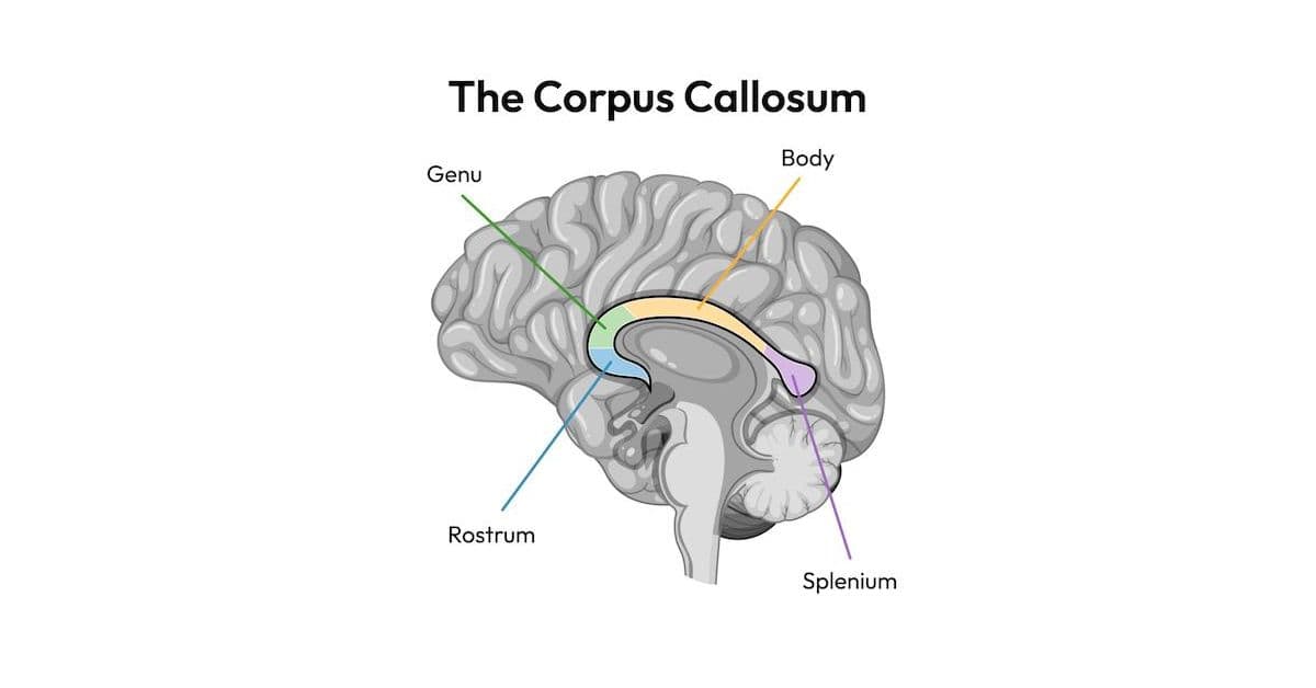
When people ask, “What is the corpus callosum?” they often want to know where it’s located as well. The corpus callosum sits deep in the center of the brain between the left and right cerebral hemispheres. It lies within the natural groove that separates the two sides of the brain, called the longitudinal fissure.
It’s shaped like a wide, curved band that stretches from the front of the brain toward the back, forming a broad “C” shape. Although it’s only about four inches (10 centimeters) long, the corpus callosum contains more than 200 million nerve fibers called axons. These fibers are covered in myelin, which gives the structure its white appearance and helps messages travel quickly between the two hemispheres.
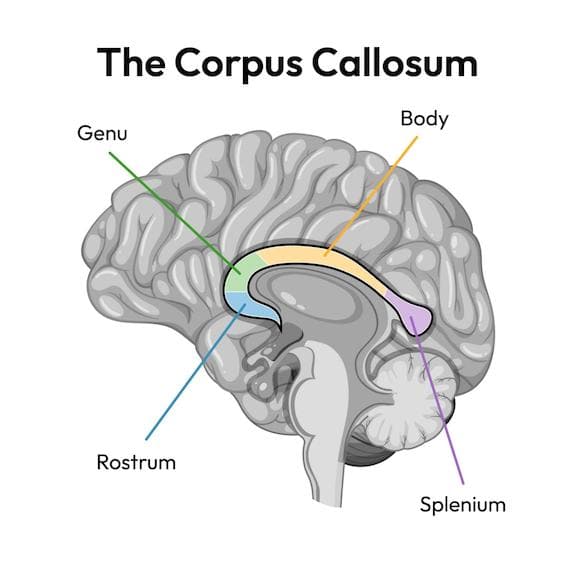
Doctors divide the corpus callosum into four regions called the rostrum, genu, body, and splenium. Each region connects different areas of the brain, allowing specialized functions to communicate with one another throughout the day.
During pregnancy, nerve fibers begin crossing between the two hemispheres around the end of the first trimester, and the corpus callosum continues developing throughout childhood as the brain matures. Doctors can clearly see the corpus callosum on an MRI scan, making medical imaging an important tool for diagnosing disorders of the corpus callosum.
What Does the Corpus Callosum Do?
The corpus callosum allows the left and right sides of the brain to exchange information almost instantly. Although each hemisphere has areas that specialize in different functions, they work together constantly. The corpus callosum helps coordinate that communication so everyday tasks feel smooth and automatic.
You rely on this teamwork more often than you might realize. For example:
- Reading involves recognizing the shapes of letters, connecting them to language, and understanding the meaning of a sentence.
- Catching a ball requires your brain to process what your eyes see, judge the ball’s speed and direction, then coordinate your muscles to react at the right moment.
- Using both hands together, whether you’re tying your shoes, chopping vegetables, or playing the piano, depends on each side of the brain sharing information so your movements stay coordinated.

The corpus callosum also helps connect brain regions involved in movement, sensory processing, learning, memory, and problem-solving.
What Happens If the Corpus Callosum Doesn’t Develop Typically?
For some people, the corpus callosum develops differently before birth. These differences are known as disorders of the corpus callosum (DCCs).
A person may be born with a completely absent corpus callosum, called agenesis of the corpus callosum (ACC). Others may have a partially formed corpus callosum, a thinner-than-expected corpus callosum, or structural differences that affect how it develops.
With DCCs, instead of relying on a single, direct pathway, the brain uses alternative routes to share information. This can affect how quickly or efficiently certain processes happen, but it also shows how adaptable the brain can be. Think of it like taking the scenic route; it might take longer, but it gets you there in a beautiful way.
Some people with DCCs experience few noticeable challenges, and others benefit from support with learning, communication, social skills, motor coordination, or other aspects of development. Many factors influence outcomes, including other brain differences and underlying genetic conditions.
The corpus callosum can also be affected later in life by conditions such as strokes, traumatic brain injuries, multiple sclerosis, tumors, or other neurological disorders. These are different from DCCs because the corpus callosum developed first and was affected afterward.
This wide range of experiences is one reason we at the NODCC focus on education, research, and connecting families with reliable information and support.
Ready to learn more? Explore our guides on each disorder of the corpus callosum, browse resources for newly diagnosed families, or connect with the NODCC community.